MRI
MRI (magnetic resonance imaging) is a technique for visualizing internal structures of the body, particularly the soft tissues. In this way, pathological alterations in the body can be detected. Inside of the MRI there is a magnet generating a magnetic field that is several thousand times higher than that of the geomagnetic field.
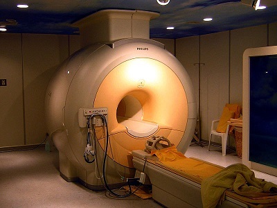
photo: KasugaHuang, license: CC BY-SA 3.0, via Wikimedia Commons
During a MRI procedure the patient is exposed a strong static magnetic field. During the MRI the magnetic characteristic of the atomic nucleus induce a certain orientation of the atoms within the magnetic field. Hereby, the atomic nuclei (e.g., hydrogen atoms) within the tissues of the body take up an identical alignment. By an electromagnetic radiofrequency pulse, this orientation is deranged. When the nuclei take back their orientation after the pulse, they emit an electromagnetic signal, which is converted to give the image.
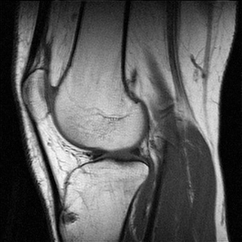
photo: Test21, license: CC BY-SA 3.0, via Wikimedia Commons
In daily clinical practice, open MRIs are normally operated from 0.2 - 1 T and closed MRIs from 1.5 - 3 T for the examination of patients. Only in research facilities, magnetic fields from 7 T up to 9.4 T are used.
Typical measurement values for MRI can be found in the database of exposure sources.